In our Cardiology Clinic we are specialized in the prevention, diagnosis and treatment of:
- Education to maintain cardiovascular health.
- Review of Cardiology. Check your cardiovascular health.
- Cardiovascular evaluation in the sportsman..
- Preoperative: Cardiological evaluation.
- Treatment of Arterial Hypertension: New! Renal Denervation treated refractory hypertension.
- Reducing cholesterol levels.
- Abandono del tabaco.
- Evaluation of chest pain
- Diagnosis and treatment and follow-up of Ischemic Heart Disease: Angina pectoris, Acute Coronary Syndrome, Myocardial Infarction.
- Diagnosis and treatment of heart valvular diseases : mitral stenosis, mitral insufficiency, aortic stenosis, aortic insufficiency, prosthetics.
- Diagnosis and treatment of palpitations: cardiac arrhythmias: Extrasystole, Atrial Fibrillation, TPSV, WPW, TRN. Ventricular arrhythmias.
- Study of Subit Death: Long QT syndrome, Brugada syndrome. Hypertrophic myocardiopathy. Arrhythmogenic Cardiomyopathy of Right Ventricle.
- Assessment and treatment of loss of consciousness:Syncope.
- Diagnosis and treatment of difficulty breathing of cardiac origin
- Diagnosis and treatment of Heart Failure.
- Evaluation of Cardiomyopathieshypertrophic, dilated, restrictive.
- Pericardial diseases: Pericarditis. Diagnosis, treatment.
- Diagnosis, treatment and follow-up of Congenital Heart Diseases: Interaricular Communication (CIA). Interventricular Communication (IVC). Persistent Arteriosus Ductus. Coartación Aorta. Complex Congenital Heart Disease.
It is important to point out the extensive experience that supports us in pediatric cardiology.
Cardiological tests
It is a test that uses sound waves to create a moving image of the heart, and with which you can measure and evaluate the flow of blood through the heart's cavities and valves.
What is it for?
It allows to study in detail all the cardiac structures (atria, ventricles, valves, etc.), and to detect if there is any anomaly in its morphology or in its operation.
It can detect abnormal blood flow inside the heart, which could indicate problems, such as an opening between the heart cavities, malfunctioning of one or more of the four heart valves, or deterioration of the heart walls. In this way, the size of the heart, its movement, or the state of its valves are accurately studied.
Types
→There are several types of Doppler echocardiography.
→Transthoracic Doppler echocardiography. Transesophageal Doppler Echocardiography
→Stress echocardiogram with stress. Pharmacological Stress Echocardiogram
→Echocardiogram with contrast. 3D Echocardiogram
→Description
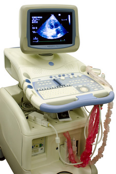
Echocardiography is a noninvasive diagnostic technique that uses ultrasound to obtain images that allow visualization of cardiac structures and large vessels.
→We can divide it into:
»Transthoracic echocardiography: if the images are obtained through the thoracic wall
»Transesophageal echocardiogram (TEE): if the images are obtained through the esophagus.
→What does it consist of?
Echocardiography uses sound waves of a certain frequency (ultrasound, sound waves that the human ear can not hear) to produce an image of the heart and see how it works. Sound waves are transmitted through the body by means of a transducer or probe, which is a device similar to a microphone; These waves bounce in the heart and return to the transducer in the form of echoes that become electrical signals that, in turn, produce images of the heart, which are seen on a screen. It is widely used in cardiology because of its availability, speed and non-invasive character.
→Types
There are different ways of processing the ultrasound signal:
»One-dimensional or M-mode echocardiography: Uses an ultrasound beam directed towards the heart; Is usually used to see only the left side (or main pumping cavity) of the heart and allows recording both the depth and the movement pattern of certain cardiac structures. Two-dimensional echocardiography or 2D mode: Produces a livelier, moving, broader image of the heart. It is one of the most important diagnostic methods.
»Doppler echocardiography: Shows how the blood circulates through the heart and large vessels, on the screen is seen in the form of colours. When focusing on the study of movement of the walls of the heart is known as tissue doppler echocardiography.
»Three-dimensional echocardiography: Provides images in three dimensions and in real time.
»Contrast echocardiography: Ultrasound contrast agents are used; Its function is to improve the echocardiographic image. Contrast is a substance that is given by injection into a vein, and when receiving the ultrasound, it is as if there were many bubbles filling the heart cavity and facilitating the distinction between the walls of the heart and the cavity.
→What is it for?
It is used to assess the functioning of muscle and heart valves, as well as to provide morphological data of other cardiac structures, such as the pericardium.
→What risks does it have?
Any.
→Possible side effects
Any.
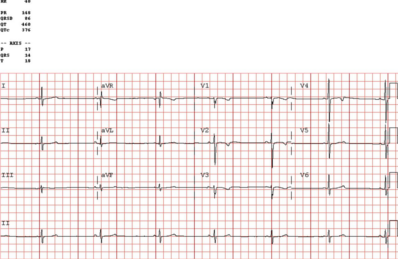 The electrocardiogram (ECG) is a study of cardiac electrical activity that serves to stimulate muscle contraction and exert the function of pumping blood.
What is it for?
→An ECG is used to measure any damage to the heart; The speed with which the heart is beating and whether it is beating normally, and the effect of drugs or devices used to control the heart. It is very useful to determine if a person has heart disease and the doctor can ask if there is chest pain or palpitations.
→It may be included as part of a routine examination in patients over 40 years of age.
→What does it consist of?
The electrocardiogram is the record in a role of the electrical activity of the heart
In a record like the photo shown in the previous tab, it is observed that on the baseline whenever a heartbeat occurs the electrical activity of the heart produces a characteristic tracing the electrocardiograph detects and graphically represents the electrical activity of the heart. It consists of a system of cables that are placed on the patient to collect the electrical activity. The cables are connected to the skin of the torso by means of adhering metallic discs called electrodes (Photo 2).
→What is it for?
The ECG is a simple test but can provide a very varied and important information. It tells us the rhythm and frequency (or number of beats or pulsations) of the heart, it can detect heart diseases, it allows to study the effect of certain medicines or detect alterations of the rest of the organism that affect the functioning of the heart. It is fundamental in the study and detection of arrhythmias.
→What risks does it have?
Under normal conditions, if the appliance and the electrical connections work properly, it is a risk-free test.
→Possible side effects
This test, under normal conditions, does not have to have side effects; In some people a redness may occur in the area where the electrodes are placed.
→Is any preparation necessary?
The patient has to lie on a stretcher wide enough to rest relaxed to avoid muscle contractures that alter the registry, and have the chest naked to place the electrodes. You have to prepare the skin and clean it well to get a good contact with the electrodes. In some cases, it is necessary to shave the hair to the patient.
→Indications
This test is recommended in the initial assessment of all patients with cardiovascular disease or to evaluate the response to treatments that may induce ECG changes. The use of serial electrocardiograms in patients with heart disease is also indicated if there are changes and even if there is no variation in cases of syncope (fainting), chest pain and extreme tiredness. Before an operation, it is considered indicated to perform an electrocardiogram to all patients with heart disease except in cases of insignificant or mild involvement, in which it would not be essential. In patients at high risk for heart disease the indications are similar. If patients remain stable and no heart disease is seen, an annual ECG is sufficient. It is considered appropriate to perform an ECG in healthy persons over 40 years of age, in patients who will be given medications that may alter the functioning of the heart or those who will be subjected to an exercise test, in case of Hard work or professions where you can risk the lives of many people. Before a surgical intervention is advised in those over 40 years and in asymptomatic diabetic patients.
→Contraindications
It is a test that has no contraindications, although in some cases it can be difficult to get a good electrocardiogram if the patient is agitated or has tremor because the muscle contraction interferes with the recording.
→Is it necessary to sign informed consent?
Do not.
What is it?
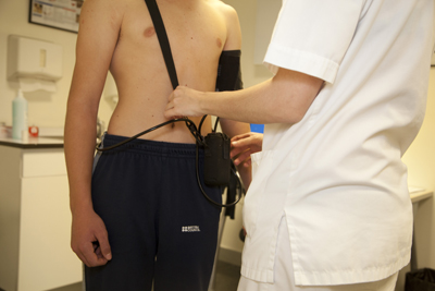
It is a test that is intended to measure a patient's blood pressure periodically, outside the medical-hospital context, over a programmable period of time, usually 24 hours a day.
The test is performed on an outpatient basis, with portable automatic devices including a cuff and the corresponding recorder. The cuff is placed on the arm and connected through a tube to the recorder. The cuff contains a sensor, which is responsible for detecting blood pressure. The recorder stores the information necessary to perform the test and the motor in charge of blowing the cuff.
What is it for?
This test is performed to measure the blood pressure of a patient, observing their variations and alterations in relation to the different phases and activities of the day. It is also used to evaluate the usefulness of antihypertensive therapy.
It is especially indicated when there is a suspicion of an increase in blood pressure induced by the act of the medical visit (effect called white coat phenomenon), that is, when there are discrepancies in the blood pressure measured in the consultation and obtained outside this.
→Other denominations
It is also known as holter of arterial pressure or by Ambulatory Blood Pressure Monitoring (ABPM).
Arterial Hypertension
It is a chronic disease characterized by higher stress levels than is considered normal. A distinction is made between systolic blood pressure (what people commonly call "discharge") greater than 140 mmHg and diastolic blood pressure ("the drop") and greater than 85 mmHg. Blood pressure is variable throughout the day and from one day to the next, so the diagnosis of arterial hypertension should be made by taking several measures separately over time, except in certain cases where a very severe elevation may be sufficient for the diagnosis.
→What does it consist of?
Repeated arterial blood pressure measurements are performed over a period of time with an automatic measurement system that stores the records in a computerized database and then analyzes those data on the computer. The patient is placed the cuff to be inflated in the flexure of the elbow of an arm. This sleeve is connected to a recording device, which is attached to the waist or with a band over the shoulder. Once the device is placed, the patient goes home and it is recommended that he live a normal life. After 24 hours, he returns to the hospital and removes the device. Then the recording device is connected to the computer and the recorded voltage measurements are read. The report can be recorded on the computer so that the doctor can later analyze it or print the result on paper.
→What is it for?
The ECG is a simple test but can provide a very varied and important information. It tells us the rhythm and frequency (or number of beats or pulsations) of the heart, it can detect heart diseases, it allows to study the effect of certain medicines or detect alterations of the rest of the organism that affect the functioning of the heart. It is fundamental in the study and detection of arrhythmias.
→Who does it?
In the case of the 24-hour voltage holter, a professional places the cuff and the recorder and prepares the system for recording. When the recording is finished, the device is removed by the nurse or the patient or a relative if they have been previously instructed. The nurse downloads the information to the computer and subsequently a doctor with appropriate training interprets the data obtained depending on the patient's clinic.
→What risks does it have?
The holter of blood pressure of 24 hours has no risk.
→Possible side effects
This test, under normal conditions, does not have to have side effects; In some people redness, may occur in the area of the cuff or in rare cases a small hematoma.
→Is any preparation necessary?
It is preferable for the patient to wear loose-fitting button-down clothing for comfort. You should keep your arm immobile when you notice that the tensioning is going to take place (the sleeve is inflated) for one minute until it is finished so that the Holter works without problems and the shots are as reliable as possible. You will not be able to shower for 24 hours and you should have a normal life.
→Indications
This is a test that is indicated in the study of variable tension figures, elevated shots, headaches...
→Is it necessary to sign informed consent?
Do not.
What is it?
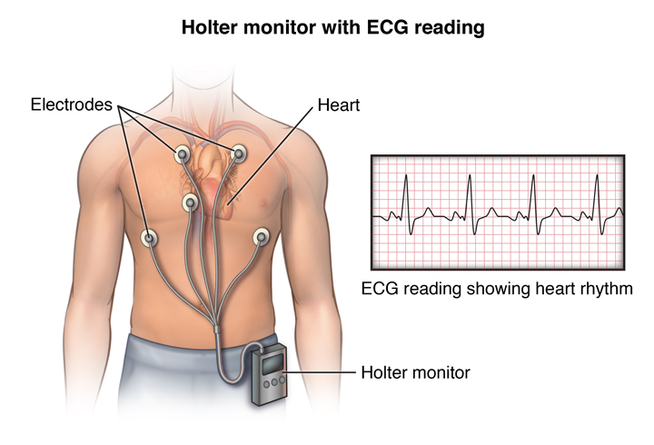
It consists of the continuous recording for 24 hours of the electrical activity of the heart, through a portable recorder that allows the patient to make his normal life during this period.
What is it for?
→To determine the origin of palpitations or symptoms of tachycardia.
→To discard slow rhythms or pauses of the heart that can lead to loss of consciousness.
→To diagnose episodes of myocardial ischemia or angina.
→To control the effectiveness of antiarrhythmic treatment in certain heart diseases.
Patient annotations tab
24-hour ECG Holter
→What is it for?
This test is useful in the diagnosis and treatment of arrhythmias, especially when the patient's symptoms or sensations can be correlated with electrocardiographic abnormalities. This test also helps assess prognosis in patients who are at high risk for arrhythmias. Was initially used to detect tachycardias (cardiac pulsations higher than normal) or bradycardia (slowing of pulsations). The technical advances have allowed that it can be used to detect parameters that suggest the possibility of dangerous arrhythmias, changes that can be associated to lack of irrigation in the heart or to alterations in the regulation of the heart rate associated to poor prognosis.
→Who does it?
In the case of continuous 24-hour holter, a professional places the electrodes and cables and prepares the system for recording. The patient with a 24-hour holter is told to note down anything he notices and the time, so he can correlate it with the record when recording data is obtained. When the recording is finished, the device is removed by the nurse or the patient or a relative if they have been previously instructed. The nurse downloads the information to the computer and subsequently a doctor with appropriate training interprets the data obtained depending on the patient's clinic.
→What risks does it have?
Continuous 24-hour holter has no risk.
→Possible side effects
This test, under normal conditions, does not have to have side effects; In some people a redness may occur in the area where the electrodes are placed.
→Is any preparation necessary?
It is preferable for the patient to wear loose-fitting button-down clothing for comfort. It is necessary to get a good adhesion of the electrodes to the skin, so it is necessary to clean the area well, and if there is a lot of hair, it may be necessary for the patient to be shaved. It is not possible to wet the device, so it is necessary to remind the patient that it is not possible to shower or bathe and it is advisable for the patient to live as normal as possible.
→Indications
This is a test that is indicated in the study of arrhythmias and in some patients with coronary risk problems and that usually manifest as palpitations, tachycardia, dizziness, fainting or chest pain.
→Contraindications
Minimal patient collaboration is required so that the electrodes are not started and the symptoms noted. It is advisable to avoid approaching electric blankets, magnets, metal detectors and high voltage areas, such as power lines, as they can affect the recording.
→Is it necessary to sign informed consent?
Do not.
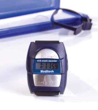
→What is it?
Innovative holter of ECG events from Meditech. The equipment is a wristwatch with two electrodes, one that is always in contact with the patient's wrist and the other on the opposite side of the watch. When the patient feels, any symptoms put the wrist of the opposite hand on the free electrode, closing the circuit and recording the ECG.
→What is it used for?
This equipment is especially suitable for cases in which in a holter of traditional ECG no pathology is detected and the patient insists on his illness.
The holter memory stores up to 15 minutes of the ECG, we can configure it from the PC to set the maximum length of the strips (do not exceed 2 min for example, even if the patient keeps the circuit closed).
We put the holter in the patient and it can stay as long as necessary until the symptoms appear. The computer warns when the memory is full so that it goes to the query to download the data.
The patient must train previously when he comes to place the clock so that the recordings that he makes are suitable and without artifacts.